Hematoxylin And Eosin Staining Of Tissue And Cell Sections

794 August 1 1998 Tissue Biology Anatomy And Physiology Histology Slides

H E Staining Kit Hematoxylin And Eosin Ab245880 Abcam

H E Staining Basics Troubleshooting Common H E Stain Problems Leica Biosystems

Figure 1 Class I Lupus Nephritis Mesangium And Capillary Loops Appear Normal In Hematoxylin And Eosin Stained Section A Ma Lupus Nephritis Lupus Microscopy

Hematoxylin Eosin Staining For Identification Of Granulomatous Download Scientific Diagram

H E Stain 02 H E Stain Stain Microbiology

Eosin is pink and stains proteins nonspecifically.
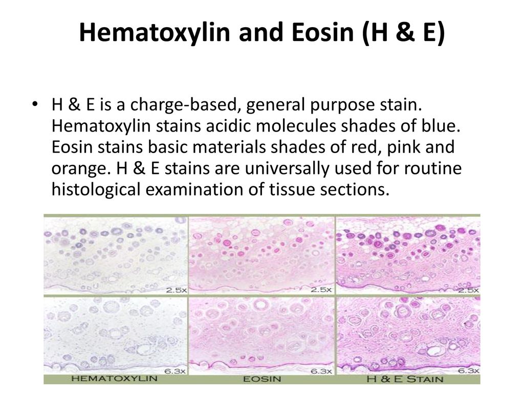
Hematoxylin and eosin staining of tissue and cell sections. In a typical tissue nuclei are. Hematoxylin has a deep blue purple color and stains nucleic acids by a complex incompletely understood reaction. The nucleus whereas eosin is the acidic dye that stains the basic components of the cell i e cytoplasm.
Fischer ah jacobson ka rose j zeller r. Hematoxylin and eosin staining of tissue and cell sections. For example when a pathologist looks at a biopsy of a suspected cancer the histological section is likely to be stained with h e.
Hematoxylin is a basic dye that stains the acidic components of the cell i e. Bahey ng abd elaziz ho gadalla kk. In a typical tissue nuclei are stained blue whereas the cytoplasm and extracellular matrix have varying degrees of pink staining.
Hematoxylin and eosin stain or haematoxylin and eosin stain is one of the principal tissue stains used in histology. Well fixed cells show considerable intranuclear detail. Hematoxylin and eosin staining technique functions to recognize different types of tissues and their morphological changes especially in cancer diagnosis.
It is the most widely used stain in medical diagnosis and is often the gold standard. Alum acts as a mordant and hematoxylin containing alum stains the nucleus light blue which turns red in the presence of acid and dark blue in the presence of alkali. Hematoxylin has a deep blue purple color and stains nucleic acids by a complex incompletely understood reaction.
Toxic effect of aflatoxin b1 and the role of recovery on the rat cerebral cortex and hippocampus. H e is the combination of two histological stains. The hematoxylin stains cell nuclei blue and eosin stains the extracellular matrix and cyto.
Histological Techniques Haematoxylin And Eosin Staining Ppt Download

Effects Of Isopropanol Storage Time On Histochemical And Immunohistochemical Stains In Lung Tissue Photomicrographs Of Lung S Red Dress Pin Science Nerd Stain
Cell Staining Protocol For Microscopy Procedures Types Techniques

Cross Section Of A Muscle Spindle Stained With Hematoxylin Eosin Located In A Young Muscle It Is Surrounded By A Capsule An Stain Photo Editing Cross Section

Blue Histology Gastrointestinal Tract Histology Slides Tissue Biology Gastrointestinal
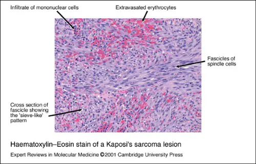
Haematoxylin Eosin Stain Of A Kaposi S Sarcoma Lesion Histologically Download Scientific Diagram

Histology Art Stained Cells Mad Scientist Science And Nature Medical Science
Hematoxylin And Eosin Staining Protocol Principle Procedure Results

Loose Connective Tissue Loose Areolar Connective Tissue Mesentery 10x Loose Connective Tissue Collagen Fibers Organic Chemistry Study

Oesophagus Histology Plate Anatomy Boutique Cells And Tissues Anatomy Human Tissue

Blue Histology Integumentary System Integumentary System Sweat Gland Medical School Motivation

Ae Practical Neural Histology Embryology Spinal Cord Pathology Study Study Of Tissues

What Histology Is And How It S Used Cells And Tissues Branches Of Science Organs
